Detecting proteins with GFP-tagging - how to visualize proteins in living cells
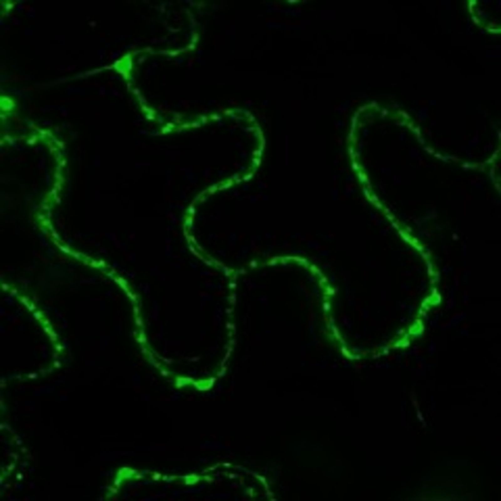
To better understand the processes inside cells, scientists have to visualize what is happening inside the living cell. Important elements of cells are proteins. They are, however, not directly visible, neither by eye nor by other means like microscopes. Therefore, the scientists couple a protein-of-interest with another fluorescent protein (here: the green fluorescent protein GFP), which can then be detected. This movie gives an overview about the general principle and the laboratory work needed for this.
Gwendolyn Kirschner, Institute for Developmental Genetics, Heinrich Heine University Düsseldorf
Planter’s Punch
Under the heading Planter’s Punch we present each month one special aspect of the CEPLAS research programme. All contributions are prepared by our young researchers.
Further Reading
Bleckmann, A., Weidtkamp-Peters, S., Seidel, C. A. M., & Simon, R. (2010). Stem cell signaling in Arabidopsis requires CRN to localize CLV2 to the plasma membrane. Plant Physiology, 152(1), 166–76. doi.org/10.1104/pp.109.149930
Somssich, M., Ma, Q., Weidtkamp-Peters, S., Stahl, Y., Felekyan, S., Bleckmann, A., … Simon, R. (2015). Real-time dynamics of peptide ligand-dependent receptor complex formation in planta. Science Signaling, 8(388), ra76. doi.org/10.1126/scisignal.aab0598
Stahl, Y., Grabowski, S., Bleckmann, A., Kühnemuth, R., Weidtkamp-Peters, S., Pinto, K. G., … Simon, R. (2013). Moderation of Arabidopsis Root Stemness by CLAVATA1 and ARABIDOPSIS CRINKLY4 Receptor Kinase Complexes. Current Biology : CB, 23, 1–10. doi.org/10.1016/j.cub.2013.01.045